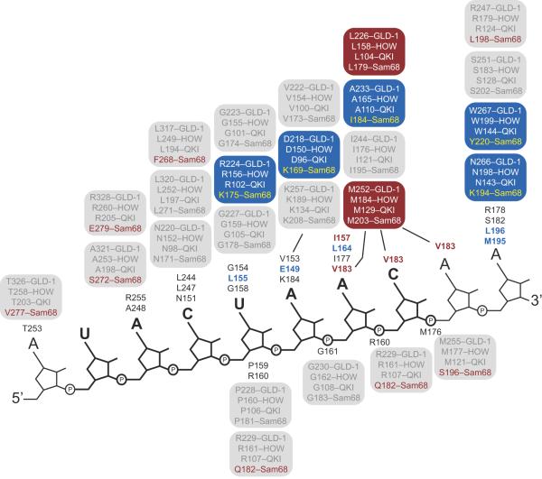
Fig. 4.
Diagram of the protein-RNA contacts observed in the SF1-BPS RNA complex structure. The sequence of the RNA is shown next to a backbone diagram. The UACUAA element is bold. The SF1 amino acids that contact RNA are presented next to the nucleotide they interact with in the structure. The corresponding amino acid in GLD-1, HOW, QKI, and Sam68 is given in a box above. Conserved amino acids are in gray. Blue boxes represent positions where all four proteins differ from SF1, but the identity of the amino acid difference is the same in GLD-1, QKI, and HOW. Red boxes represent positions where all four proteins differ from SF1 but the difference is identical in all four STAR proteins. Red font indicates a position where only Sam68 differs from the SF1 sequence.

