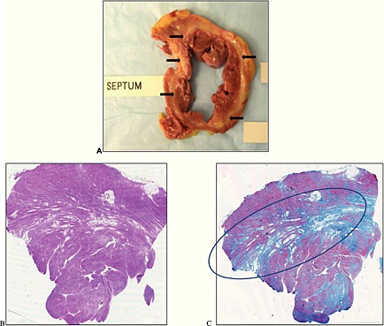
Figure 1.

Cross‐section of explanted heart, H&E, and Masson's trichrome stain. Panel A shows a cross‐sectional of the native heart with a near circumferential midwall fibro‐lipomatous degeneration of the left ventricle (arrows). Panel B represents the microscopic examination of the cross‐section depicted in panel A with haematoxylin and eosin stain. This shows hypertrophic myocytes with areas of fibrosis with predilection in the midwall (circle) as evidenced by Masson's trichrome staining in Panel C.
