Figure 3.

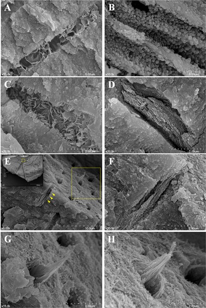
A–D: Diverse nanocrystallographs of pozzolan‐based (Pz‐) MTA sealer cement induced intratubular biomineralization. A: Organized nanoflakes (×10,000), (B) microsphere (×10,000), (C) mixed (×20,000), or (D) organized plates (×20000) are intergrown and exploited to seal the dentinal tubules. (E‐F) Successive intratubular biomineralization of gutta‐percha and Pz‐MTA sealer filled canals. E: Boxed area (yellow) of left upper lower magnification image (×35) from the horizontal split specimen obturated with gutta‐percha and Pz‐MTA sealer cement showing the interface at 350–400 μm distance from dentinal tubule orifice (×5,000). (F) The pre‐crystallization seeds observed along the collagen fiber (arrowheads of (E), ×20,000). (G, H) A higher magnification of upper right boxed area of (E) showing successive intratubular biomineralization in either (G) plate‐like form or (H) agglomeration of precipitation seeds along the collagen fibrils (×20,000). Scanning electron microscope images.
