
Figure 1.
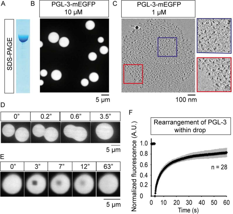
PGL-3 forms liquid-like drops in vitro.
(A) SDS-PAGE of PGL-3-mEGFP expressed and purified from insect cells.
(B) Maximum intensity projection of series of confocal z- slices shows PGL-3-mEGFP at 10 μM phase-separates into drops.
(C) Virtual slice from cryo-electron tomography, 5 nm thick, shows a drop of PGL-3-mEGFP (1 μM). Red and blue boxes show zoomed in view.
(D) Time-lapse single confocal plane micrographs show two PGL-3-mEGFP drops fuse with each other into a single drop within 3.5 s.
(E) Time-lapse single confocal plane micrographs show fluorescence recovery of PGL-3-mEGFP after an internal 1.44 μm × 1.44 μm area is photobleached at 3 s.
(F) Quantification of FRAP data presented in (E), n = 28, error bars represent 1 SD.
See also Figure S1, Movies S1–S2.