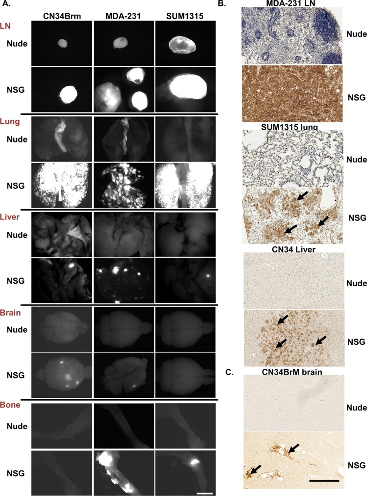
Fig 2. Human breast cancer cells are more metastatic in NSG mice than in nude mice in a spontaneous metastasis model.
Metastases were analyzed from the mice described in Fig 1. A. Fluorescent images (white area is fluorescent tumors) were taken using Zeiss SteroDiscovery.V12 fluorescence dissecting microscope with an AxioCam MRm digital camera. Representative images are shown from several different organs. Bar, 2 mm. B. To confirm the metastases, breast cancer cells were identified in the lymph nodes (LN), lung, and liver using a HLA antibody and in the brain using a human specific cytokeratin 7 antibody. Arrow denotes tumor area. Bar, in B is 1 mm.

