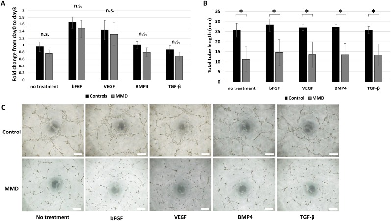
Fig 3. Cellular functions of iPSCs-derived endothelial cells (iPSECs).
Panels A shows the results of CC8 cell proliferation assay (A) after 3 days of culture. Panel B shows that representative photomicrographs of the tube formation assay. Panel C shows the quantitative analysis of total tube length. *: P < 0.05, error bars: SD, n.s.: No significance. Scale bar: 500 μm.

