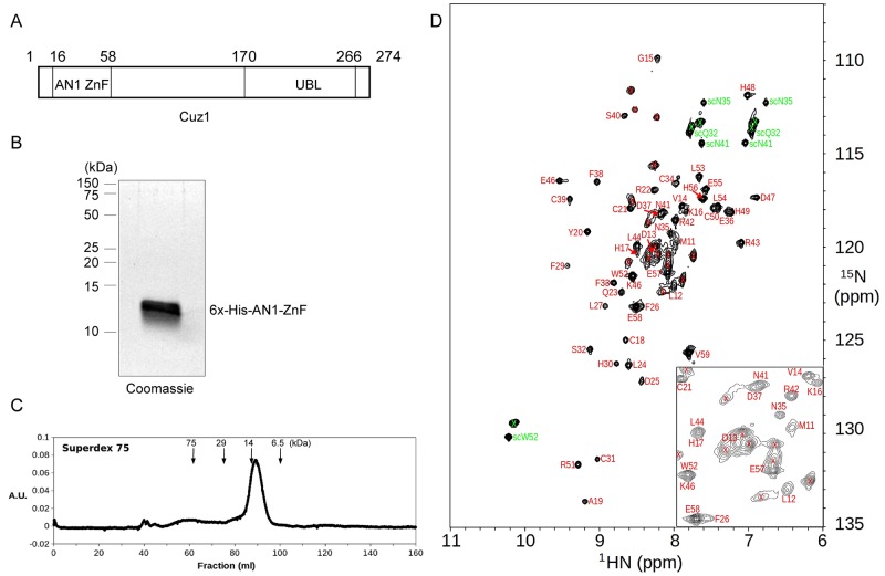
Fig 1. Expression and Purification of the Cuz1 AN1 Zinc Finger Domain.
A) Schematic diagram of the Cuz1 protein. UBL, ubiquitin-like domain. B) Purified Cuz1 AN1 ZnF protein (15 μg) as analyzed by SDS-PAGE followed by Coomassie staining. C) Size exclusion chromatography of purified Cuz1 AN1 ZnF protein. Molecular weight size standards are shown for reference. D) 15N-HSQC spectrum of the 15N-labeled Cuz1 AN1 ZnF domain sample showing assignments of Cuz1 residue resonance peaks. Backbone amide and sidechain peaks are labeled in red and green, respectively, and peaks from the cloning tag are marked with a “x” sign. An expanded view of the central spectral region is shown at the lower right corner.

