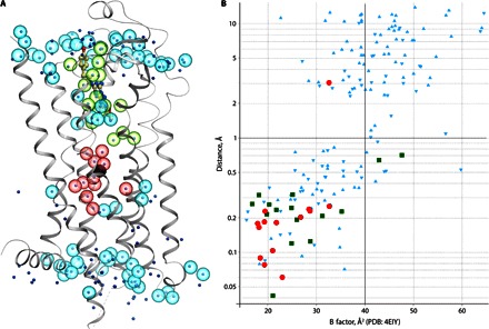
Fig. 3. Comparison of resolved water molecules between the room temperature XFEL structure (A2A_S-SAD_1.9) and the cryocooled synchrotron structure (PDB: 4EIY).

(A) Cartoon representation of the XFEL structure with overlaid waters. Water molecules from the XFEL structure are shown as semitransparent spheres, whereas waters from PDB: 4EYI are shown as dots, colored by location: green, close proximity to ligand (<5 Å); red, sodium ion pocket (<10 Å); cyan, other regions. (B) Conservation of the water positions between PDB: 4EIY and XFEL structures. For each water molecule in PDB: 4EIY, the distance to the closest water in the XFEL structure is shown on the y axis, whereas its B factor is shown on the x axis. Data points are colored the same way as in (A). Positions of water molecules can be considered as conserved if the distance between corresponding water molecules in two structures is less than 1 Å.
