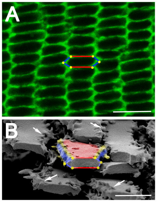
Figure 2.
Lens fiber cells have a hexagonal geometry when viewed in cross section. (A) An equatorial cross section of a mouse lens stained with fluorescent wheat germ and viewed with a confocal microscope. The hexagonal geometry of cells is highlighted in the center of the image showing the two broad sides of the cell labeled in red, the four short sides labeled in blue and the six vertices represented as yellow dots. Scale bar= 7.5μm. (B) A scanning electron micrograph showing lens fiber cells cut across their major axis allowing for viewing of their hexagonal geometry. The yellow vertices and white arrows show that there are membrane protrusions seen along these edges. Red– broad side; blue– short side; yellow– vertices/membrane protrusions; Scale bar= 5μm

