Figure 3.

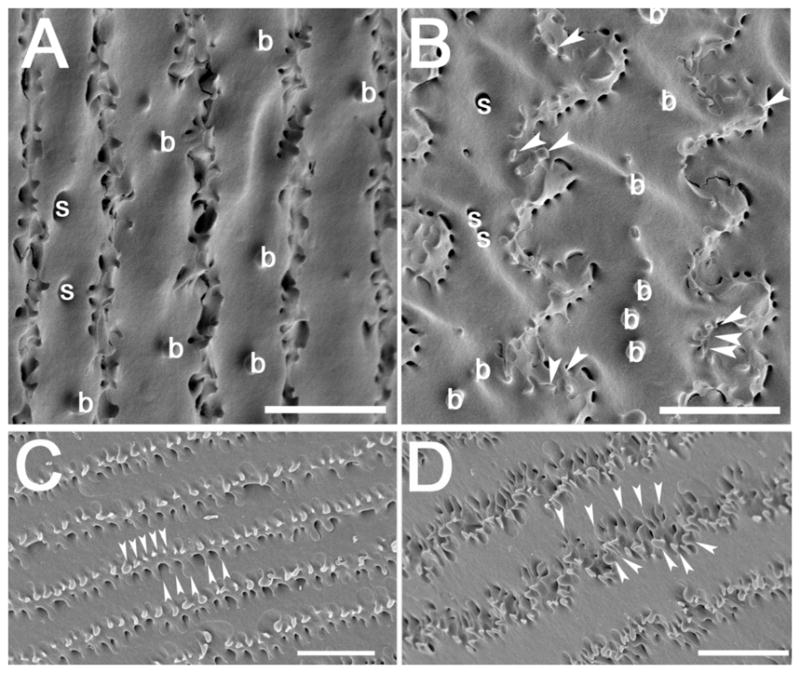
Scanning electron micrographs showing ball and sockets on newly differentiated fiber cells and the formation of elaborate membrane protrusions in mouse lenses. (A) An electron micrograph showing straight fiber cells dotted with ball and sockets along the broad side of the cell in the youngest lens fiber cell layers. (B) As fiber cells mature, ball and sockets are seen along the broad sides of cells, and membrane protrusions (arrowheads) are first seen along the vertices of these cells. C) Deeper in the lens cortex, morphologically obvious ball and socket junctions are not seen, but the membrane protrusions become more obvious (arrowheads) D) In the deepest layers of the lens cortex, the membrane protrusions become even more elaborate (arrowheads). arrowheads- membrane protrusions; b– ball; s– socket; Scale bar for all panels= 5μm.
