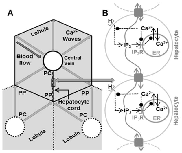
Figure 1.
A – Hepatocyte cords and Ca2+ wave propagation in liver lobules. At the microscopic scale, the liver consists of roughly hexagonal shaped lobules. The area surrounding the central vein is the pericentral region (PC), whereas the outer extremities are the periportal (PP) region. Blood flows from the PP region towards the PC region in lobules. Hepatocytes lie roughly radially between the PC and PP regions along quasi-linear structures in the hepatic plate. Each hepatocyte is connected to its neighbors by gap junctions. Ca2+ waves originate near the PC region and propagate radially outward along all hepatocyte cords. B - Network model of Ca2+ wave propagation along hepatocytes in a sinusoid induced by the agonist.

