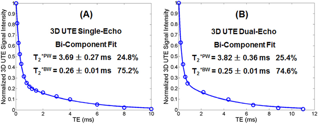
Figure 3.
Bi-component analysis of 3D UTE images of a bovine cortical bone sample acquired with the single-echo protocol (A) and dual-echo protocol (B). Bone images from the single-echo protocol show a bi-component T2* decay with a short T2*BW of 0.27 ± 0.01 ms and a longer T2*PW of 2.37 ± 0.36 ms, accounting for 73.4% and 26.6%, respectively. Bone images from the dual-echo protocol show a bi-component T2* decay with a short T2*BW of 0.27 ± 0.01 ms and a longer T2*PW of 2.26 ± 0.17 ms, accounting for 70.5% and 29.5%, respectively.

