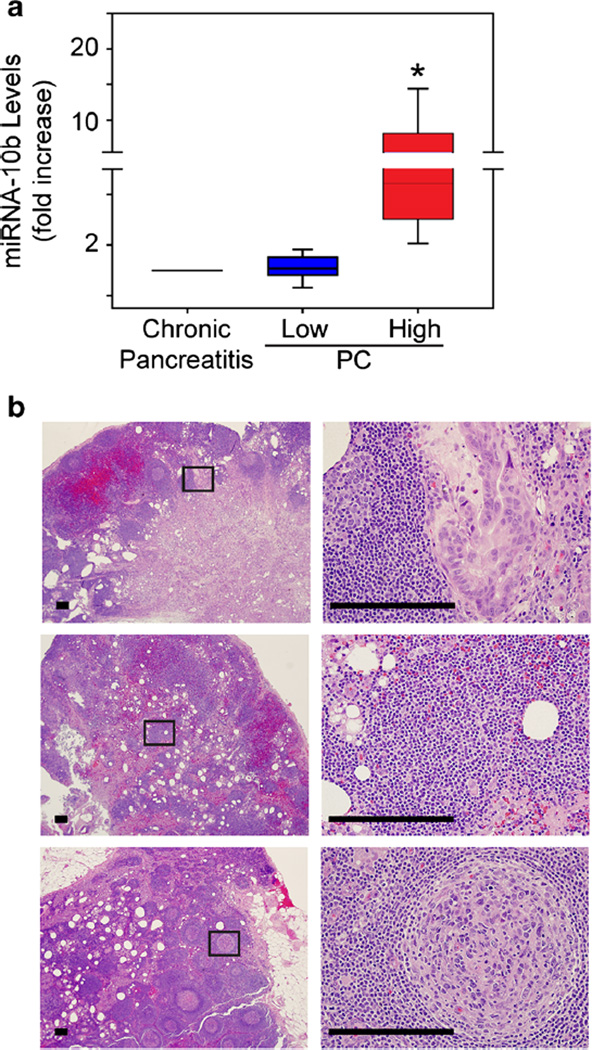
Fig. 1.
Elevated miRNA-10b levels in a subset of PC station 8 nodes. a Quantitative PCR for miRNA-10b in station 8 lymph nodes from chronic pancreatitis or PC patients shows that this miRNA is significantly increased in nodes from some PC patients (high, red bar; *p < 0.05), whereas other nodes have levels that are similar to chronic pancreatitis (low, blue bar). Data are presented as interquartile range (IQR). b H&E staining of station 8 lymph nodes from PC patients with high miRNA-10b. Shown are representative images from a non-recurrent patient with moderately differentiated PC (top) and recurrent patients with poorly differentiated PC (middle) or without cancer cells (bottom) in the station 8 lymph node. Panels on the right are magnified images of boxed areas. Scale bars, 50 µm

