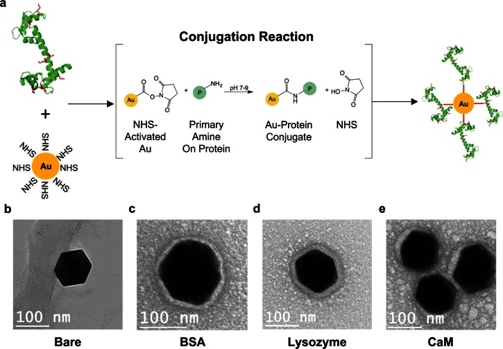
FIG. 2.
Nanoparticle bioconjugation. (a) Proteins, in this schematic CaM, are introduced to 100 nm NHS-activated AuNPs where the NHS on the particle and primary amine on surface lysines of the protein (in red) react to form covalent protein-conjugated nanoparticles. (b)–(e) Transmission Electron Microscopy (TEM) images of bare, BSA, lysozyme, and CaM conjugated AuNPs, respectively. The AuNP itself appears as the larger dark object in the image. The protein corona can be seen as a halo-like feature around the particle circumference.

