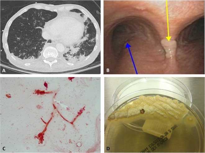
FIG 1.
(A) CT of the patient's chest showing nodular infiltrates highly suggestive of fungal infection. (B) Bronchoscopy with a fungal growth (yellow arrow) on the carina, next to the right main stem bronchus (blue arrow). (C) Direct Gram stain of patient's tracheal aspirate showing tapering phialides with phialoconidia suggestive of Penicillium or Paecilomyces species. (D) Yellow-tan colonies on Sabouraud dextrose agar characteristic of Paecilomyces species.

