Fig. 1.
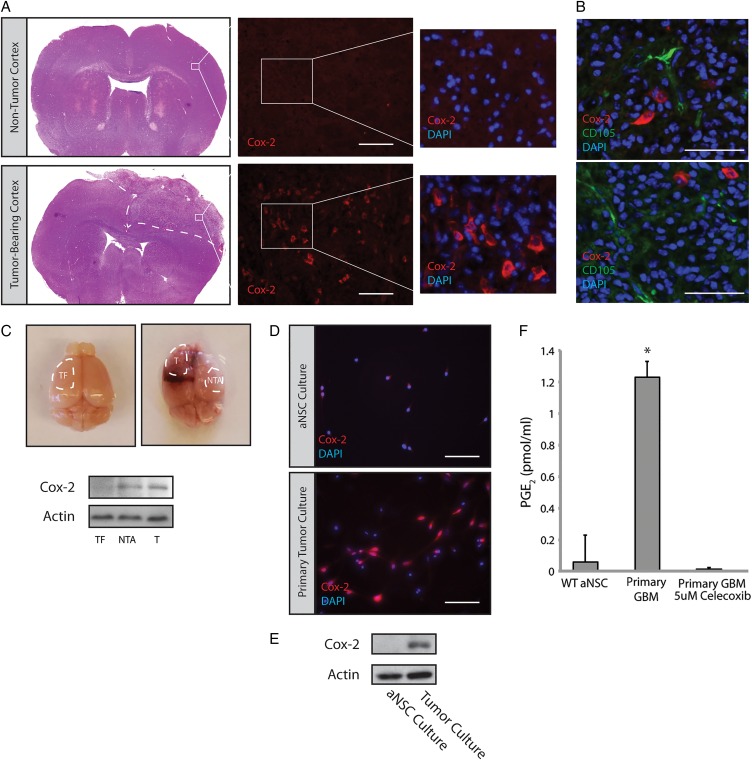
Cox-2 is overexpressed in glioblastoma (GBM)-bearing mouse brains. (A) GBMs generated via injection of PDGFB-expressing virus into the lateral ventricle of Nestin-tva, Arf −/− mice showed significant expression of Cox-2 compared with a corresponding cortical region in a nontumor-bearing mouse. Scale bar = 0.2 mm. (B) Double-labeling with Cox-2 and the tumor vasculature marker CD105 showed minimal overlap. Scale bar = 0.1 mm. (C) Tumor (T) and Nontumor adjacent (NTA) tissue were isolated from tumor-bearing brains (n = 3) and analyzed by Western blot. Cox-2 was consistently elevated in T and NTA tissue relative to tumor-free (TF) tissue. (D) Dissociated GBMs were used to generate adherent primary tumor cultures. These cultures continued to express Cox-2 by immunofluorescence staining. Scale bar = 0.1 mm. (E) Cox-2 protein levels were determined by Western blot using lysates from primary cultures of GBM versus adult neural stem cells (aNSC). (F) PGE2 levels were analyzed by ELISA on conditioned media from primary aNSC and GBM cultures treated with 5 µM celecoxib or vehicle for 24 hours. Error bars represent mean ± SEM (n = 3). *, P < 0.05.

