
Figure 1. Cellular crosstalk among autophagic pathways and neutral lipases.
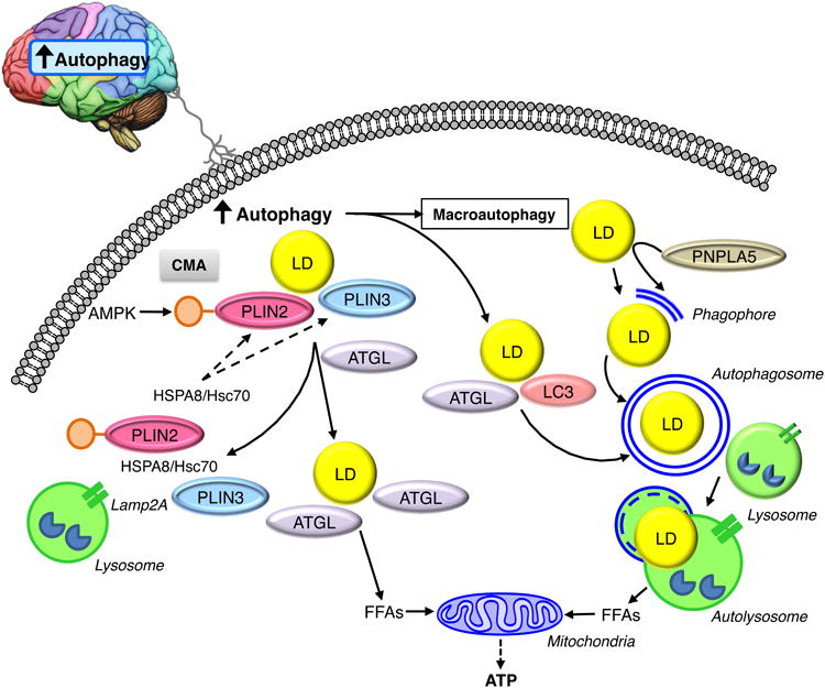
Autophagy in the brain sends signals that induce lipophagy by promoting the interactions of LC3 and ATGL on the LD surface, and by increasing the direct removal of LDs by macroautophagy. PNPLA5 may promote autophagy by supplying lipids from LDs necessary for the formation of the pre-autophagosome membrane structure the phagophore. CMA promotes neutral lipolysis by degrading AMPK-phosphorylated PLIN2 and PLIN3, thereby allowing ATGL access to the lipid core of the LD. Together lipophagy and neutral lipolysis generate FFAs that drive mitochondrial FFA β-oxidation to supply the cell with ATP.