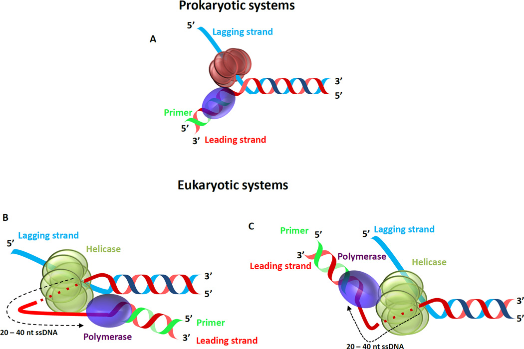
Figure 1. Helicase - DNA polymerase at the replication fork.
(A) Cartoon showing the hexameric ring shaped replicative helicase and the replicative DNA polymerase enzymes bound to a replication fork DNA that represents an intermediate structure during leading strand replication. (B & C) Two proposed models for the architecture of the eukaryotic replisome based on single particle EM studies [15]. Depending on the orientation of the helicase with respect to the fork junction (If the C-terminal domain of the helicase is facing the fork junction the leading strand DNAP is present ahead as in (B) or if the N-terminal domain of the helicase is facing the fork junction the leading strand DNAP is behind the helicase as in (C). Further studies that capture the DNA path are required to identify the correct orientation of the system.

