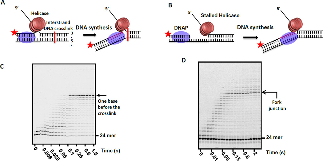
Figure 7. Mapping the position of the DNAP active site on the replication fork with the helicase.
(A) DNA substrate with an interstrand transplatin DNA crosslink positioned approximately in the middle of the dsDNA region of the fork substrate. Extension of the labeled primer by the helicase-DNAP complex is carried out in the presence of all dNTPs to map the position of the DNAP active site with respect to the transplatin crosslink. (B) DNA substrate with 20-nt ssDNA gap between the primer end and fork junction. T7 helicase is stalled at the fork junction using dTMP-PCP, a non-hydrolysable dTTP analog, and the extension of the primer by the DNAP to the point of the stalled helicase is followed using a labeled primer. (C) High resolution sequencing gel shows the time course of primer extension. T7 DNAP is able to reach all the way to the crosslink point. (D) High resolution sequencing gel shows the time course of the extension of the primer up to the fork junction without stalling at any upstream positions.

