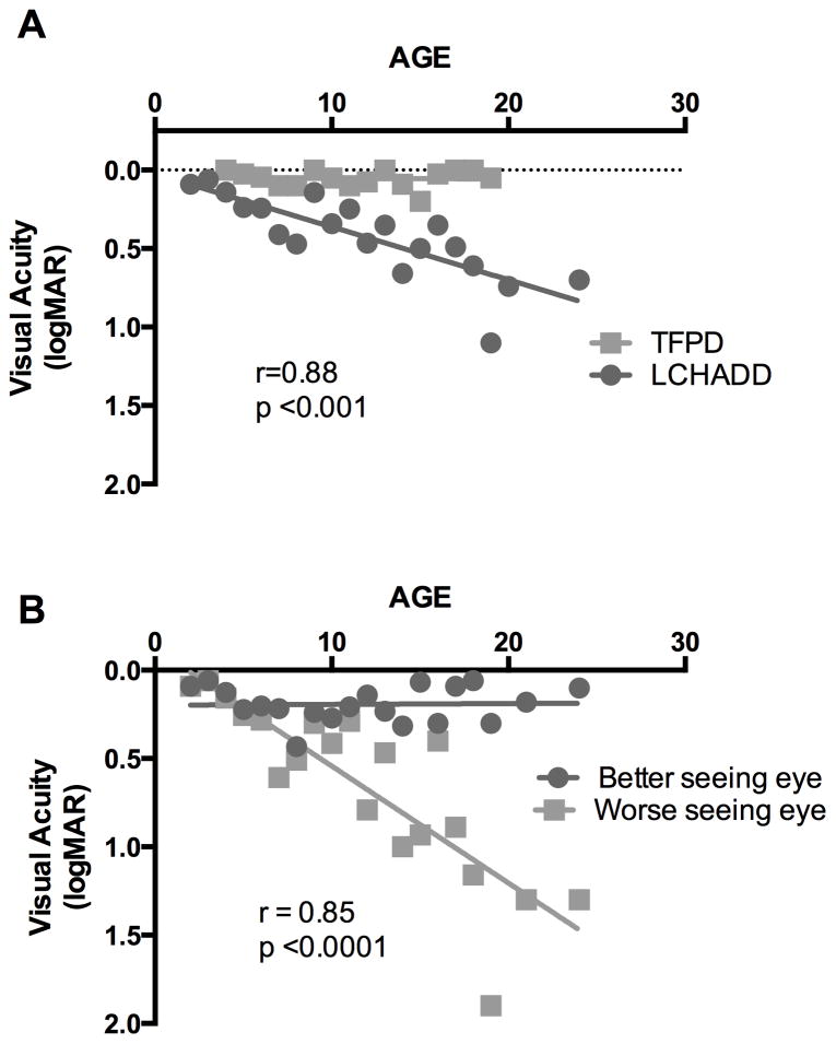
Figure 1. Visual acuity declines in LCHADD eyes but not TFPD eyes.
(A) Subjects with LCHADD (circles) had a progressive decline in best-corrected visual acuity with increasing age when all visits of all patients were averaged for each age rounded to nearest year, averaging 0.03 loss of logMAR acuity per year. The data was non-normally distributed. Spearman correlation between logMAR and age was significant (r=0.88, p<0.001). TFPD patients (squares) maintained excellent visual acuity throughout the study. There was no significant correlation between logMAR and age among subjects with TFPD. (B) Best-corrected visual acuity plotted individually for the better (circles) and worse seeing (squares) eye in LCHADD patients. There was a significant correlation between logMAR and age for the worse seeing eye among subjects with LCHADD (Pearson r= 0.85; p<0.0001).

