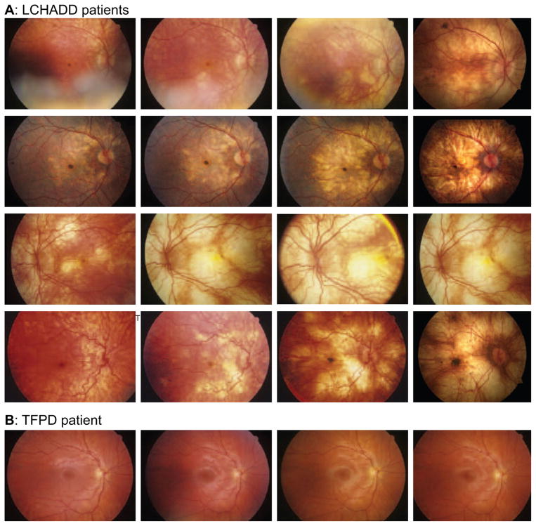
Figure 4.
(A) Color fundus photos demonstrate disease progression despite dietary modification and systemic disease control in several patients with LCHADD. Supratitles in each row indicate the patient and identified genetic mutations. Early in the disease, there is an accumulation of pigment at the central macula, which is followed by a progressive patchy chorioretinal atrophy of the posterior pole. With time, the atrophy extends to involve the periphery. In some subjects, there is relative sparing of peripapillary retina. Late stages of the disease show the development of a subfoveal pigmentary scar (not in all patients). There is minimal retinal vascular attenuation or disc pallor. (B) Color fundus photos demonstrate disease stability in one patient with TFPD. Supratitles indicate the patient and identified genetic mutations. Unlike the LCHADD patients, subjects with TFPD did not have any evidence of pigment clumping or chorioretinal atrophy. The fundus photos of subject LC16 remained normal over 4.1 years of photographic follow up. Although not included within the figure, the remaining two subjects with TFPD also did not have any notable changes on fundus photography.

