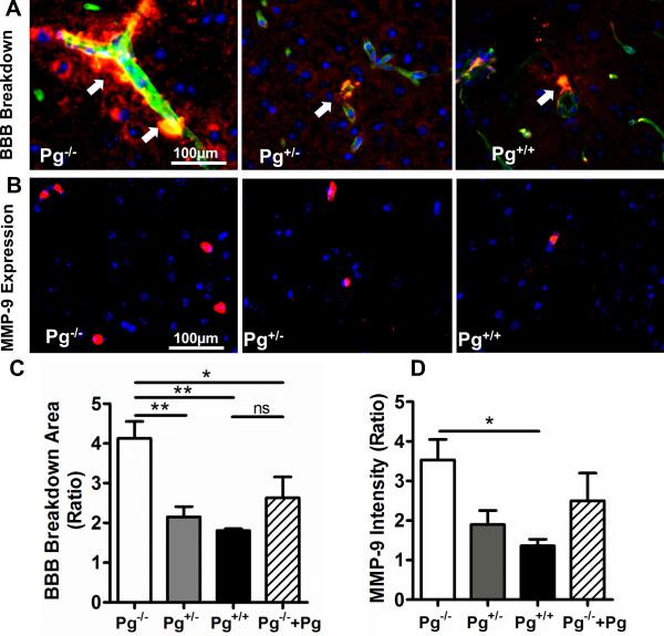
Figure 3. Higher Pg levels are associated with decreased BBB breakdown and reduced vascular MMP-9 expression after stroke.
(A) Leakage (arrows) of albumin (red) into the brain parenchyma outside collagen IV-stained (green) blood vessels was assessed in the ischemic hemispheres by comparison to the non-ischemic hemispheres of Pg−/−, Pg+/− and Pg+/+ mice. Merged images (20x) show areas of overlapping staining for collagen IV and albumin (yellow) and DAPI-stained nuclei in blue. (B) Representative immunofluorescence images (20x) showing MMP-9 expression (red) in the ischemic hemispheres of Pg−/−, Pg+/− and Pg−/− mice as detected by goat anti-mouse MMP-9 primary antibody followed by donkey anti-goat DyLight® 549. (C) The bar graphs show the ratio of the total area of albumin staining (in 20x fields) and (D) MMP-9 fluorescence intensity in the ischemic vs. the non-ischemic hemisphere of each mice group (mean ± SE). n= 4-5 per group, *p<0.05, **p<0.01, ns (not significant).

