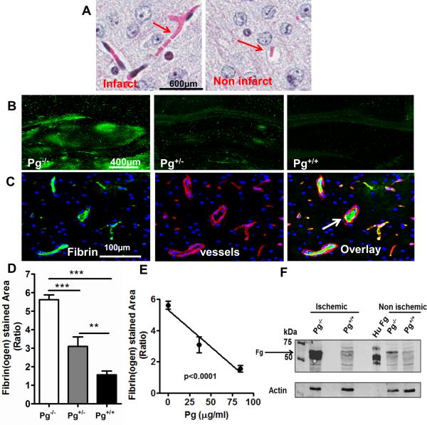
Figure 4. Higher plasma Pg levels are associated with reduced microvascular thrombosis in the ischemic brain.
(A) MSB stained brain images showing microvascular thrombosis (pink fibrin) in the ischemic and non-ischemic hemisphere (B) Representative immunofluorescence images (6x) of the hippocampal region of ischemic hemispheres of Pg−/−, Pg+/−, and Pg+/+ mice showing areas of fibrin(ogen) deposition (green). Rabbit anti-mouse fibrin(ogen) and Dylight® 488 donkey anti-rabbit were used as primary and secondary antibodies respectively. (C) Representative brain images from Pg−/− mice (20x) showing fibrino(gen) (green), collagen IV-stained blood vessels (red) and a merged image showing intravascular fibrin(ogen) (white arrow) in blood vessels. For fibrin(ogen) and blood vessels, rabbit anti-mouse fibrin(ogen) and goat anti-human collagen IV were used as primary antibodies respectively. Dylight® 488 donkey anti rabbit and Alexa Fluor® 555 donkey anti-goat respectively were used as secondary antibodies. (D) Quantitation by digital imaging of the fibrino(gen) stained area in 15 different fields at 20x magnification and expressed in the bar graph as ratio of ischemic vs non-ischemic hemispheres of Pg−/−, Pg+/−, and Pg+/+ mice. Mean ± SE, n= 5 per group. (E) Regression analysis (line graph) shows that fibrin(ogen) deposition in brain was inversely correlated to Pg levels. (F) Immunoblotting of brain samples with rabbit anti-mouse fibrino(gen) antibody showed fibrin(ogen (α,β chains; Fg) in the ischemic and non-ischemic hemisphere. Purified human fibrinogen (Hu Fg) was used as a positive control.

