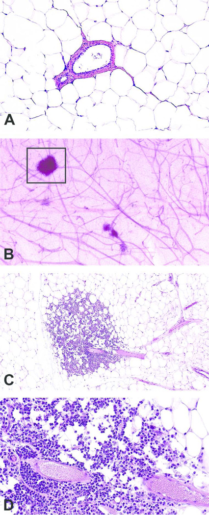
Figure 2. Mammary gland section of perivascular inflammation using the whole mount method.
Normal mammary gland section with no histopathologic findings, formalin-fixed H&E-stained, magnification 10x (A); Increased opacity around ducts and stromal structures (box) in the contralateral carmine-stained mammary whole mount (B); At low magnification, in the contralateral mammary gland H&E prepared from the whole mount (boxed area shown), there are clusters of mononuclear cells around the blood vessel and extending into the adjacent adipose tissue magnification 20x (C); Higher magnification illustrated that the inflammation is composed of perivascular lymphocytes; magnification 40x (D). Tissue samples are from a 14 mo. old female CD-1 mouse.

