Fig. 1.
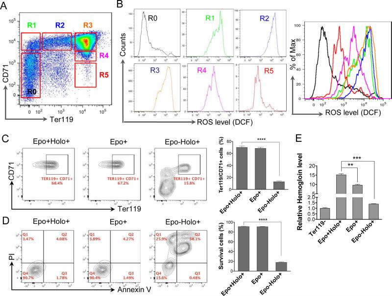
Erythropoietin and transferrin regulate the generation of reactive oxygen species during mouse fetal liver terminal erythropoiesis. (A) Flow cytometric analysis of E13.5 mouse fetal liver cells stained with CD71 and Ter119. R0 to R5 populations were gated based on the cells’ relative expression levels of CD71 and Ter119. (B) Flow cytometric analysis of the levels of reactive oxygen species (ROS) based on the intensity of CM-H2DCFDA in R0-R5. The colors of the curves (right) match the gated populations (left). (C-D) Flow cytometric analysis of differentiation (C) and apoptosis (D) of cultured mouse fetal liver erythroblasts based on the Ter119 and CD71 level, and annexin V and propidium iodine (PI), respectively. Ter119 negative fetal liver erythroblasts were purified and cultured in SCF medium for 8 h followed by 24 h culture in indicated medium. Statistical analysis with student's t-test is on the right. Data were obtained from three independent experiments and presented as mean ± SD. **** p <0.0001. Epo: Erythropoietin; Holo: holo-transferrin. (E) Relative hemoglobin levels from cells in C. Equal number of the cells were collected. Total hemoglobin was quantified by measuring light absorbance at 540 nm using Drabkin's reagent. ** p <0.01, *** p <0.001.

