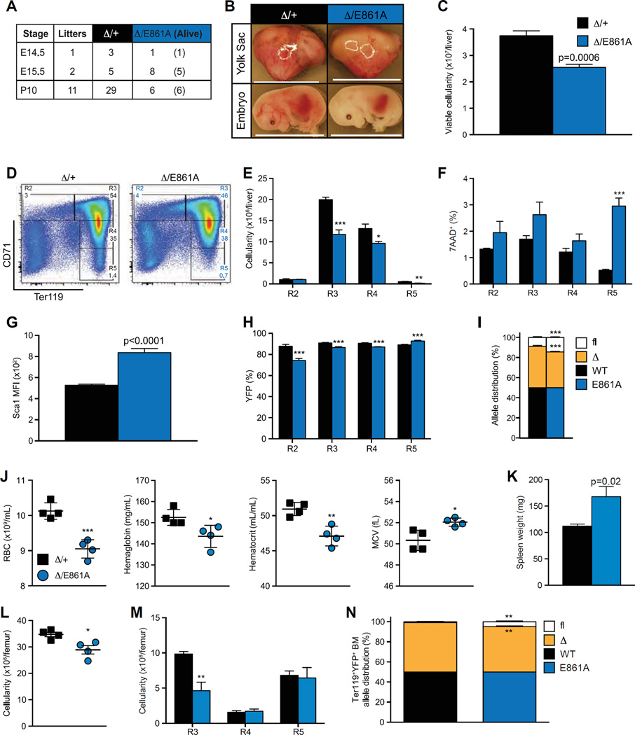
Figure 5.
A-to-I RNA editing by ADAR1 is required for fetal erythropoiesis. (A) Survival of EpoR-Adar1Δ/E861A (Δ/E861A) embryos compared with EpoR-Adar1Δ/+ littermate controls (Δ/+). (B) Representative images at E15.5. Scale bar: 1.2 cm. (C-H) FL analysis of Δ/E861A and Δ/+ at E15.5. (C) Total viable (7-AAD−) FL cellularity. (D) Representative FACS plots. (E) Enumeration of erythroid cells. Frequency of (F) dead (7-AAD+) and (G) YFP+ R2-R5 erythroid cells. (H) Sca1 MFI. (I) Allele distribution of Adar1flAdar1Δ, and Adar1+ or Adar1E861A of FL cells at E15.5–17.5 as determined by gDNA semi-qPCR. (J–N) Analysis of ≥ 16-week-old Δ/E861A and Δ/+ mice. (J) PB RBC counts, hematocrit, and hemoglobin levels and mean RBC volume. (K) Spleen weights. Cellularity of whole BM (L) and erythroid fractions (M). (N) Adar1 allele distribution of FACs isolated Ter1 19+YFP+ BM. Results are mean ± SD (E15.5, n = 5; E16.5, D/+ n = 2 and Δ/ E861A n = 1; E17.5, Δ/+ n = 1 and Δ/E861A n = 2; ≥ 16 week, n = 4). *p< 0.05, **p< 0.005, and ***p< 0.0005 compared with Δ/+.

