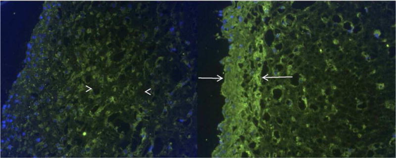
Figure 6. Immunohistochemical labeling for type I collagen in fibrin-ASC tissue constructs.

Bilayered construct on the left shows green collagen labeling in the middle segment (arrowheads). Homogeneous construct on the right shows intense labeling near the surface (arrows). In both, nuclei are labeled blue. (Original magnification: 20×)[97] Reprint with permission from Long, J. L.; Neubauer, J.; Zhang, Z.; et al. Otolaryngol.- Head Neck Surg. 2010, 142, 438. Copyright (2010) SAGE Publishing.
