
Figure 7. H&E stained tissue slides of HA-Dextran crosslinked hydrogel implanted in the ferret vocal fold at week 3.
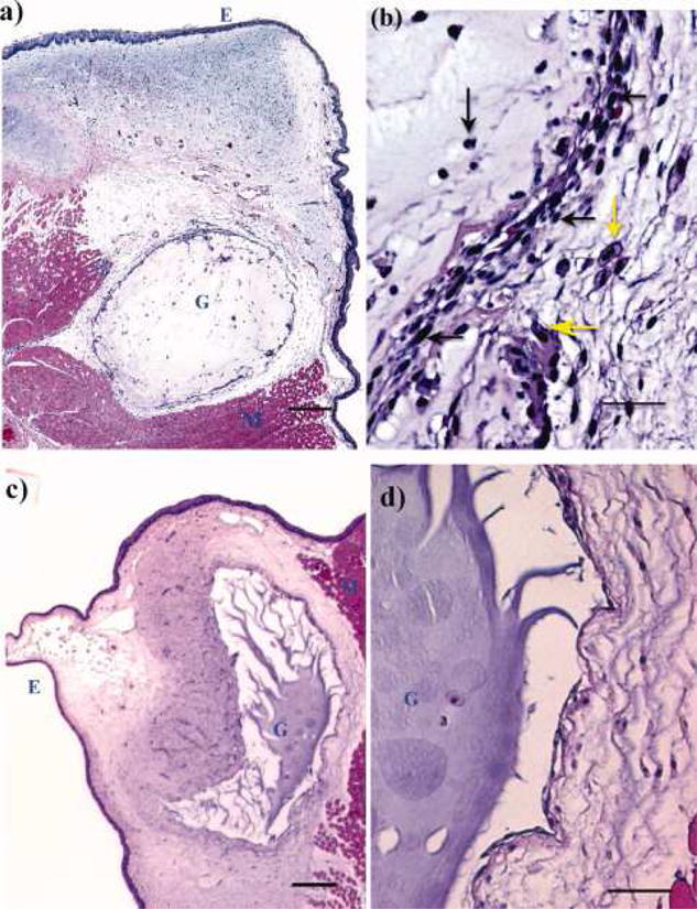
Regions of epithelium, injected gel and muscle tissue are indicated by letters E, G, and M, respectively. (a) HA-dex0.75 in the host tissue (scale bar: 200μm); (b), the interface of the host tissue and HA-dex0.75 gel (scale bar: 50μm); (c), HA-dex1 in the host tissue (scale bar: 200μm); (d) the interface of the host tissue and HA-dex1 gel (scale bar: 50μm). Cell types were visually identified in b and pointed by arrows (yellow horizontal arrow: macrophage; yellow vertical arrow: foam cell; black vertical arrow: neutrophil; black horizontal arrows: fibroblasts).[113] Reprint with permission from Luo, Y.; Kobler, J. B.; Heaton, J. T.; et al. J. Biomed. Mater. Res. B. Appl. Biomater. 2010, 93, 386. Copyright (2010) Wiley.