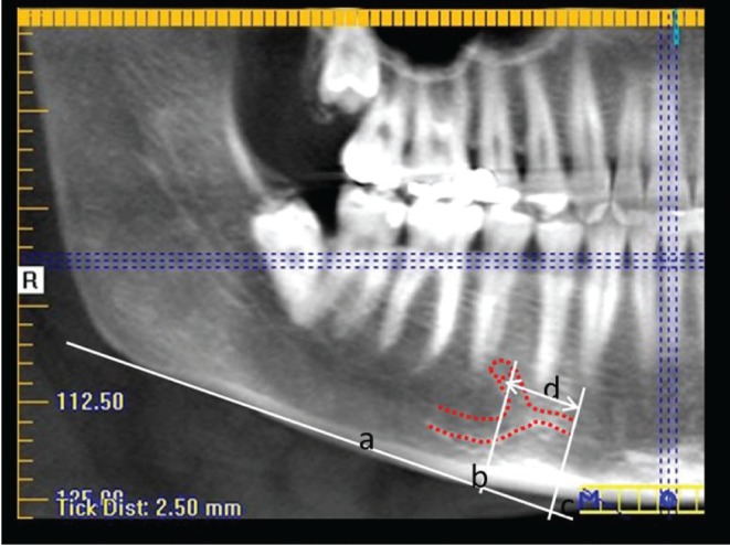
Fig. 2. Visualization of the anterior loop and incisive canal (red dotted line) on the panoramic reconstructions. a: The lower mandibular cortex as the plane of reference. b: The line perpendicular to the line passing through the mesial border of the mental foramen. c: The line perpendicular to the line passing through the most mesial point of the anterior loop of the mental nerve and the mandibular incisive canal. d: The distance between lines b and c, corresponding to the mesial length of the extent of the anterior loop or incisive canal.

