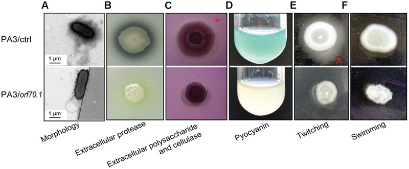
FIGURE 7.

Phenotypic analyses of P. aeruginosa (PA3/ctrl and PA3/orf70.1). (A) Transmission electron microscope image. (B,C) Detection of extracellular protease, polysaccharide and cellulase of P. aeruginosa on LB plates containing 2% milk or 0.1 congo red. (D) Pyocyanin of P. aeruginosa in LB medium. (E,F) Motility experiments on LB plates with 1% agar for twitching and 0.3% agar for swimming.
