. 2016 Mar 12;13(2):e20919. doi: 10.5812/iranjradiol.20919

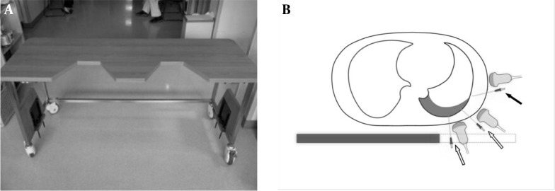
Figure 1. A, A table with gaps at one side of the tabletop was constructed for the cadaver experiment. With the lateral margin of the table carved out, the posterior thorax was exposed for the ultrasound probe and tapping needle to approach it from below. B, Drawing of axial cross section in a typical supine patient with a small-to-moderate effusion. Compared to the conventional method of lateral approach (solid arrow), the effusion can be much more easily visualized and tapped from the posterior and posterolateral aspects of the hemithorax (open arrows) via the carved out (dashed line) portion of the novel supine thoracentesis tabulation.

Figure 1.