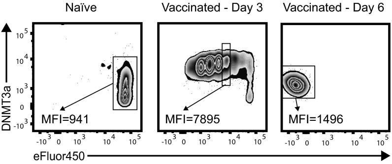
Fig. S4.
Kinetics of DNMT3a expression after activation. WT OT1 CD8+ T cells were adoptively transferred into congenic hosts. Splenocytes were isolated 3 or 6 d after infection with VacOva. Representative plots are gated on CD8+Thy1.1+ T cells from uninfected (Left) or VacOva-infected (Middle and Right) mice. MFI, DNMT3a mean fluorescence intensity in transferred OT1 T cells that were either naïve and undivided, in the third division 3 d postinfection, or had undergone multiple divisions 6 d postinfection. n = 3 mice per group. This experiment was performed three times with similar results.

