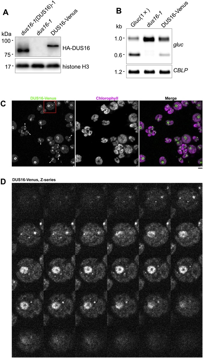
Fig. S5.
Complementation of miR9897-mediated silencing in dus16-1 by expression of DUS16-Venus and additional images of DUS16-Venus localization. (A) Immunoblotting of DUS16 tagged with an anti-HA antibody. Strain DUS16-Venus #1 expresses Venus-HA-FLAG-tagged DUS16 in a dus16-1 background. Each panel is representative of three independent experiments. (B) Northern blotting of gluc mRNA in the indicated strains. Each panel is representative of three independent experiments. (C) A field of dus16-1 cells expressing DUS16-Venus. Maximum-projection images of z-stacks covering the cell bodies are shown. The arrowheads indicate eyespot autofluorescence in some of the cells. (Scale bar, 5 µm.) (D) Montage of z-stacks of the area indicated by the red square in C, showing DUS16-Venus localization in the nucleus and as cytoplasmic punctae. The same cell also was used to generate a set of average-projection images shown in Fig. 2.

