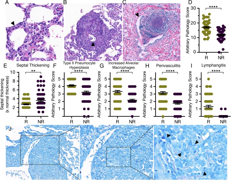
Fig. 4.
SIV-induced pathology and the presence of bacilli perivascular lesions. H&E staining of lung sections from Mtb/SIV coinfected reactivators and nonreactivators displayed exacerbated pathology, including (A) interstitial pneumonia with septal thickening (white arrowhead), type II pneumocyte hyperplasia (black arrowhead), and lymphohistiocytic infiltration; (B) perivasculitis (black arrowhead showing blood vessel wall); and (C) lymphangitis (black arrowhead showing lymphatic vessel membrane). (D–I) Multiple lung sections from reactivators and nonreactivators were scored in a single-blinded fashion by a board-certified veterinary pathologist and quantitatively compared as (D) total SIV-induced pathology, (E) septal thickening, (F) type II pneumocyte hyperplasia, (G) increased alveolar macrophages, (H) perivasculitis, and (I) lymphangitis. Samples from three animals in each group were analyzed, with 12 lung sections analyzed per animal. Each dot corresponds to a section analyzed. **P < 0.01; ****P < 0.0001 using Student’s t test. (J) Ziehl–Neelsen staining revealed the presence of numerous intact, rod-like tubercle bacilli, indicated by black arrowheads in a perivascular lesion in a reactivator. Right panel is a magnified image of the boxed region from Left and Center panels. (Magnification, A, 50×; B, C, and J, 40×.)

