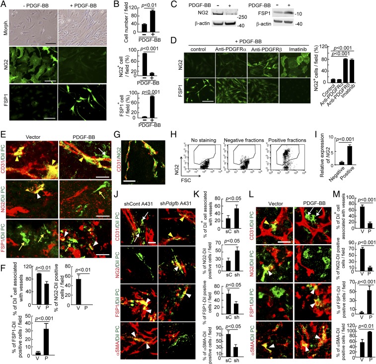
Fig. 2.
PDGF-BB induces PFT in vivo and in vitro. (A) Morphology, NG2 positivity, and FSP1 positivity in PDGF-BB–stimulated and nonstimulated primary pericytes. (Scale bar, 100 μm.) (B) Quantification of cell proliferation, NG2+ signals, and FSP1+ signals (n = 8 fields per group). The experiments were repeated three times. (C) Western blot analysis of NG2 and FSP1 proteins in PDGF-BB–stimulated and nonstimulated primary pericytes. β-actin detection indicates the standard loading in each lane. (D) NG2+ signals and FSP1+ signals in buffer-, anti-PDGFRα-, anti-PDGFRβ-, and imatinib-treated pericytes that were stimulated with PDGF-BB. Quantification of NG2+ cells in various treated groups (n = 8 fields per group). The experiments were repeated two times. (Scale bar, 100 μm.) (E) DiI-labeled pericytes (green) were implanted into T241-vector and T241-PDGF-BB tumors. Tumor vessels were stained with CD31 (red in Top), pericytes were stained with NG2 (red in Middle), and fibroblasts were stained with FSP1 (red in Bottom). Yellow arrowheads indicate vessel-associated DiI-labeled pericytes. White arrows indicate vessel-disassociated injected pericytes. The white arrowhead in the middle panel points to NG2 and DiI double signals and the yellow arrow in the middle panel indicates NG2− Dil+ cells. Yellow arrows in the lower panels indicate FSP1− Dil+ cells and white arrowheads in the lower panels point to FSP1+ Dil+ cells. (Scale bars, 25 μm.) (F) Quantification of CD31+ vessel-associated DiI+pericytes, NG2+DiI+ structures, and FSP1+DiI+ structures (n = 12 fields per group). Data are represented as mean ± SEM. P, PDGF-BB; V, vector. (G) Double immunohistochemical staining of human cancer tissue with CD31 (red) and NG2 (green). (Scale bar, 50 μm.) (H) FACS analysis of isolated NG2+ human pericytes from human tumors. (I) Quantification of NG2 positive signals of isolated human pericytes by quantitative PCR (triplicates per sample). (J) Tracing DiI-labeled human pericytes (green) implanted in scrambled shRNA- and shPdgfb-transfected human A431 tumors. White arrows indicate vessel-disassociated pericytes, yellow arrowheads point to vessel associated pericytes, and white arrowheads indicate double positive signals. (Scale bar, 25 μm.) (K) Quantification of CD31+vessel-associated DiI+pericytes, NG2+DiI+ structures, FSP1+DiI+ structures, and αSMA+DiI+ structures (n = 10 fields per group). Data are represented as mean ± SEM. sC, scrambled control tumor; sh, shPdgfb-transfected tumor. (L) Tracing DiI-labeled human pericytes (green) implanted in vector- and PDGF-BB-LLC tumors. The yellow arrowhead points to vasculature-associated pericytes, and white arrows indicate vasculature-disassociated pericytes. White arrowheads indicate double positive signals. (Scale bar, 25 μm.) (M) Quantification of CD31+vessel-associated DiI+pericytes, NG2+DiI+ structures, FSP1+DiI+ structures, and αSMA+DiI+ structures (n = 10 fields per group). Data are represented as mean ± SEM. P, PDGF-BB; V, vector.

