Fig. 4.
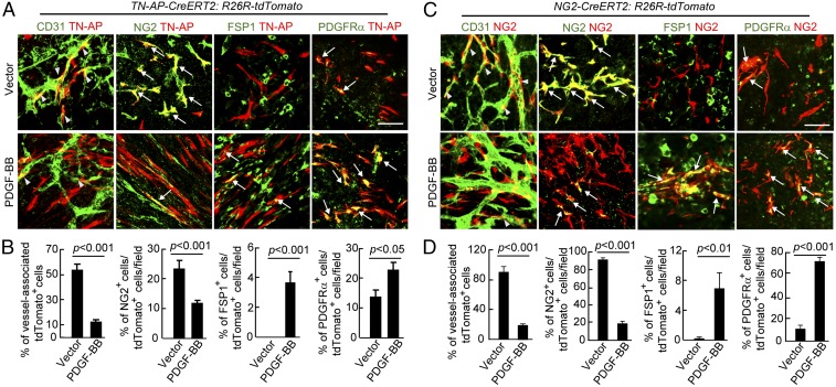
Genetic tracing of pericytes in contribution to PFT. (A) Genetic tracing of differentiation of alkaline phosphatase (AP)-marked pericytes into fibroblasts in T241-vector and T241-PDGF-BB tumors. Tamoxifen-induced AP-tdTomato-red (red) tumor tissues were contained with CD31, NG2, FSP1, and PDGFRα. Yellow arrows indicate overlapping positive signals in each panel. Arrowheads point to vessel-associated AP-tdTomato+ pericytes. (Scale bar, 50 μm.) (B) Quantification of vessel-associated AP-tdTomato+ cells, the total number of NG2+ AP-tdTomato+ structures, FSP1+ AP-tdTomato+ structures, and PDGFRα+ AP-tdTomato+ structures (n = 15 fields per group) in T241-vector and T241-PDGF-BB tumors. (C) Genetic tracing of differentiation of NG2-marked pericytes into fibroblasts in T241-vector and T241-PDGF-BB tumors. Tamoxifen-induced NG2-tdTomato-red (red) tumor tissues were contained with CD31, NG2, FSP1, and PDGFRα. Yellow arrows indicate overlapping positive signals in each panel. Arrowheads point to vessel-associated NG2-tdTomato+ pericytes. (Scale bar, 50 μm.) (D) Quantification of vessel-associated NG2-tdTomato+ cells, the total number of NG2+ NG2-tdTomato+ structures, FSP1+ NG2-tdTomato+ structures, and PDGFRα+ NG2-tdTomato+ structures (n = 15 fields per group) in T241-vector and T241-PDGF-BB tumors. Data are represented as mean ± SEM (n = 4 animals per group).

