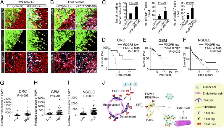
Fig. 6.
PDGF-BB–stimulated pericytes promote primary cancer invasiveness. (A) Invasiveness of nonstimulated pericyte- EGFP+ T241-vector cell- and PDGF-BB–stimulated pericyte- EGFP+ T241-vector-coimplanted primary tumors in relation to FSP1+ stromal fibroblasts (white) and CD31+ intra- and peritumoral microvessels (red). Dashed lines mark the tumor rims and arrows point to the invasive front. n = 8 animals per group. (Scale bar, 50 μm.) (B) Invasiveness of nonstimulated pericyte- EGFP+ T241-vector cell- and PDGF-BB–stimulated pericyte- EGFP+ T241-vector-coimplanted primary tumors in relation to αSMA+ (white) stromal myofibroblasts and CD31+ intra- and peritumoral microvessels (red). Dashed lines mark the tumor rims and arrows point to the invasive front. n = 8 animals per group. (Scale bar, 50 μm.) (C) Quantification of invasive tumor cells, numbers of FSP1+ stromal fibroblasts, and numbers of αSMA+ stromal myofibroblasts at the invasive fronts of nonstimulated pericyte-T241-vector cell- and PDGF-BB–stimulated pericyte-T241-vector-coimplanted primary tumors (n = 10 fields per group). (D) Kaplan–Meier survival of PDGFB-high (n = 36) and PDGFB-low (n = 36) COADREAD patients. (E) Kaplan–Meier survival of PDGFB-high (n = 107) and PDGFB-low (n = 105) LGG patients. (F) Kaplan–Meier survival of PDGFB-high (n = 199) and PDGFB-low (n = 192) LUSC patients. (G) Correlation of FSP1 expression in PDGFB-high (n = 36) and PDGFB-low (n = 36) COADREAD patients. (H) Correlation of FSP1 expression in PDGF-BB-high (n = 107) and PDGF-BB-low (n = 105) LGG patients. (I) Correlation of FSP1 expression in PDGFB-high (n = 199) and PDGFB-low (n = 192) LUSC patients. (J) Diagram of tumor-derived PDGF-BB in promoting tumor invasion and metastasis. Tumor cell-derived PDGF-BB builds up a high gradient that attracts pericytes to move away from tumor microvessels. Once pericytes detach, they differentiate into stromal fibroblast (PFT) in the presence of PDGF-BB. Increases of stromal fibroblasts and myofibroblasts in tumors significantly promote tumor cell invasion and intravasation into the circulation, leading to an increase of CTCs and distal metastasis. Data are represented as mean ± SEM.

