Fig. S2.
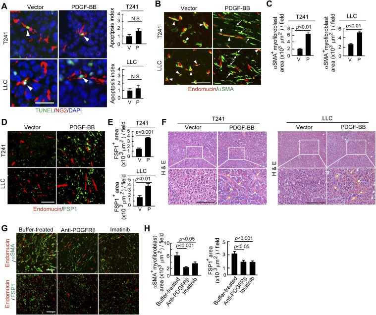
Gain of PDGF-BB function in mouse tumors increases stromal fibroblasts and myofibroblasts and ablates pericytes. (A) TUNEL immunostaining of apoptotic cells in T241-vector and -PDGF-BB and LLC-vector and -PDGF-BB tumors. NG2+ and TUNEL+ apoptotic cells were quantified (n = 30 fields per group). P, PDGF-BB; V, vector. (Scale bar, 25 μm.) (B) Endomucin+ vessels (red) and αSMA+ signals (green) in T241-vector and -PDGF-BB and LLC-vector- and -PDGF-BB tumors. Yellow color indicates the overlapping signals (arrowheads). Arrows point to vessel-disassociated αSMA+ signals. (Scale bar, 100 μm.) (C) Quantification of αSMA+ myofibroblasts in T241-vector and -PDGF-BB and LLC-vector- and -PDGF-BB tumors (n = 12 fields per group). P, PDGF-BB; V, vector. (D) Endomucin+ vessels (red) and FSP1+ signals (green) in T241-vector and -PDGF-BB and LLC-vector- and -PDGF-BB tumors. (Scale bar, 100 μm.) (E) Quantification of FSP1+ fibroblasts in T241-vector and -PDGF-BB and LLC-vector- and -PDGF-BB tumors. (n = 12 fields per group). P, PDGF-BB; V, vector. (F) H&E staining of T241-vector and -PDGF-BB tumor tissues. Arrows indicate the tumor stromal components. (Scale bar, 100 μm.) (G) (Upper) Endomucin+ tumor vessels (red) and αSMA+ cells (green). (Lower) Endomucin+ tumor vessels (red) and FSP1+ stromal fibroblasts (green) in buffer-, anti-PDGFRβ-, and imatinib-treated T241-PDGF-BB tumors. (Scale bar, 100 μm.) (H) Quantification of αSMA+ myofibroblsts (n = 12 fields per group) and FSP1+ fibroblasts (n = 12 fields per group) in buffer-, anti-PDGFRβ-, and imatinib-treated T24-PDGF-BB tumors. Data are represented as mean ± SEM.

