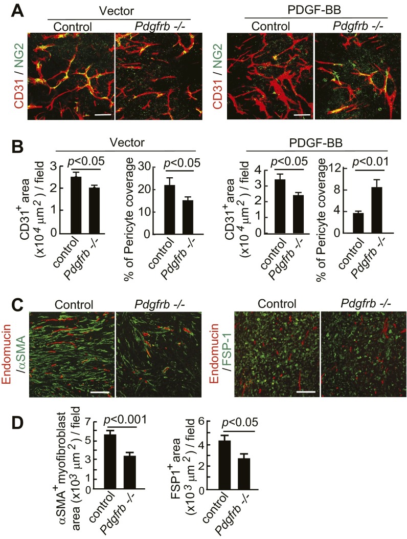
Fig. S5.
Deletion of Pdgfrb in mice ablates PDGF-BB–stimulated stromal components. (A) CD31+ (red) vessels and NG2+ pericytes (green) in T241-vector and T241-PDGF-BB tumors grown in Pdgfrbflox/flox mice (control mice) and Pdgfrb−/− mice. (Scale bar, 50 μm.) (B) Quantification of CD31+ microvessel density and pericyte coverage in T241-vector and T241-PDGF-BB tumors grown in Pdgfrbflox/flox mice (control mice) and Pdgfrb−/− mice. (n = 7 fields per group). (C) Endomucin+ (red) vessels and αSMA+ signals (green) and endomucin+ (red) vessels and FSP1+ signals (green) in T241-PDGF-BB tumors grown in Pdgfrbflox/flox mice (control mice) and Pdgfrb−/− mice. (Scale bar, 100 μm.) (D) Quantification of αSMA+ myofibroblasts and FSP1+ fibroblasts in T241-PDGF-BB tumors grown in Pdgfrbflox/flox mice (control mice) and Pdgfrb−/− mice. (n = 12 fields per group). Data are represented as mean ± SEM.

