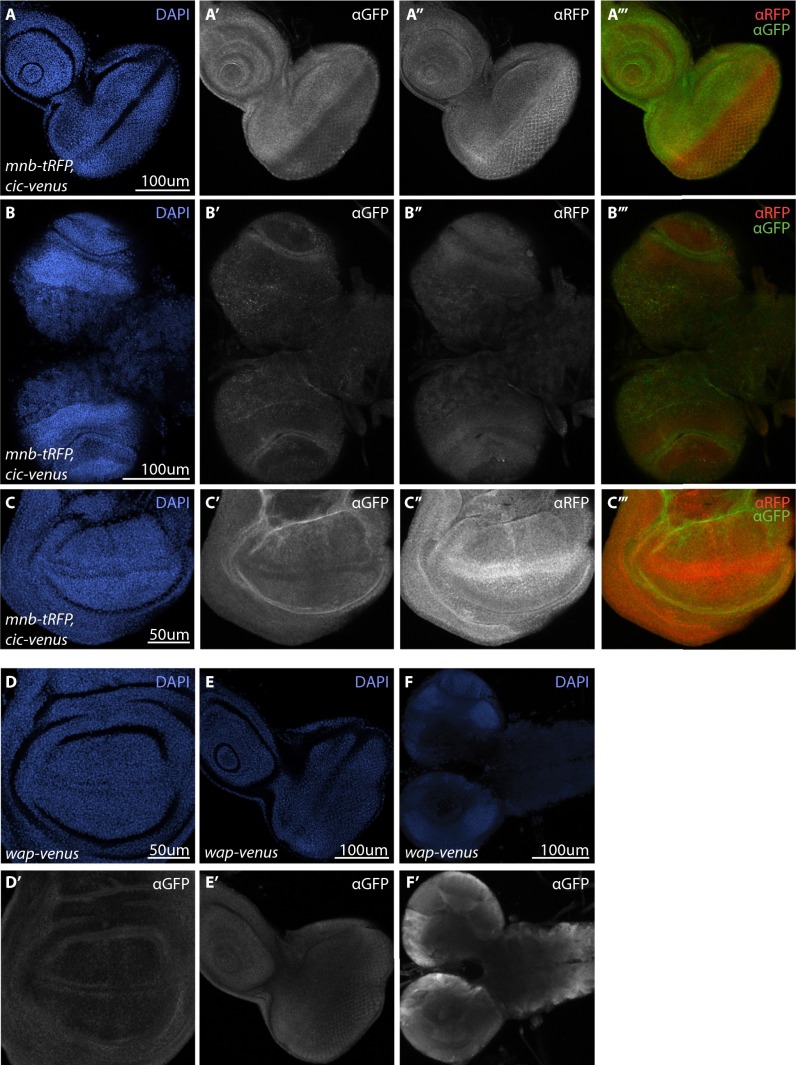
Fig. S2.
Mnb-RFP, Cic-Venus, and Wap-Venus expression in third instar larval tissues. (A–F) Cic-Venus and Wap-Venus expression was detected by GFP antibody, Mnb-tRFP was detected by tRFP antibody and cell nuclei were detected by DAPI (blue). (A′–C′′′) Cic-Venus and Mnb-tRFP expression in eye imaginal discs (A–A’’’), brain optic lobes (B’–B’’’), and the pouch region of wing imaginal discs (C’–C’’’). For merged panels (A’’’–C’’’), Cic-Venus is in green and Mnb-tRFP is in red. (D–F’) Wap-Venus expression (gray) in wing imaginal discs (D’), eye imaginal discs (E’) and larval brain (F’). (Scale bars: A–B′′′, E, and F, 100 µm; C and D, 50 μm.)

