Fig. 5.
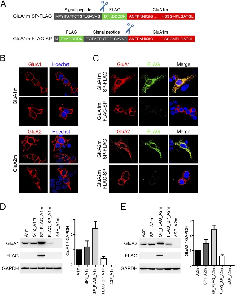
SPs were cleaved off after AMPAR translation. (A) Schematic representation of the constructs. The FLAG tag (green box) was inserted either behind the amino-terminal SP (black box) or behind the starting methionine residue. (B) Representative immunofluorescence staining for GluA1 (red) and GluA2 (red) in transfected HEK 293T cells with a GluA1m or GluA2m construct, respectively. Nuclei were stained with Hoechst 33258 (blue). (C) Representative immunofluorescence staining for GluA1 (red) and FLAG (green) in transfected HEK 293T cells with GluA1m SP-FLAG or GluA1m FLAG-SP construct designed in A, respectively. Nuclei were stained with Hoechst 33258 (blue). (D and E) Relative expression level of GluA1 (D) and GluA2 (E) when manipulations were carried out around SP.

