Fig. 3.
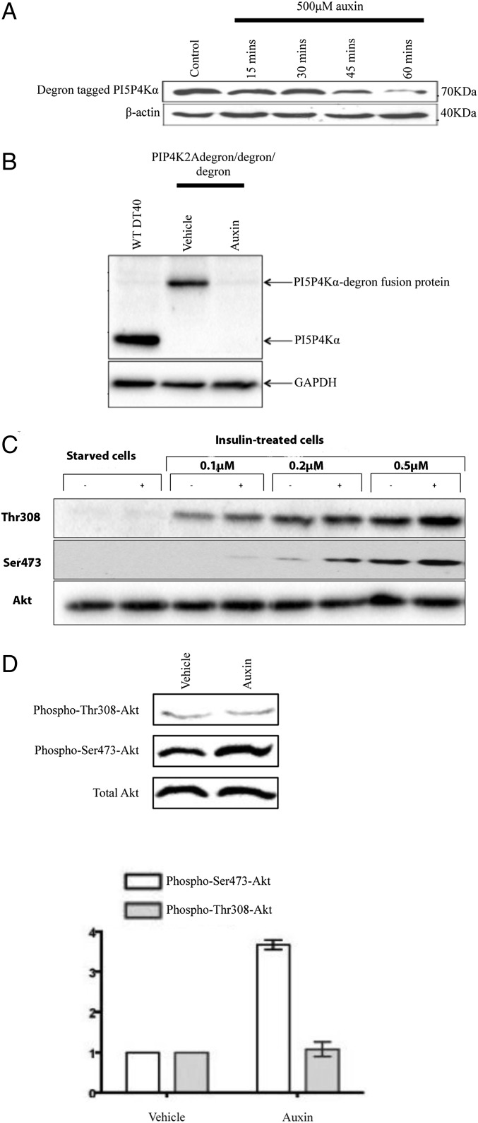
Acute removal of PI5P4Kα with the auxin degron system. (A) Cells (PIP4K2Adegron/degron/degron) were treated with auxin as indicated, and any remaining PI5P4Kα degron fusion protein was immunoprecipitated against its FLAG tag and blotted against its poly-His tag. (B) Whole-cell lysates blotted with an anti-PI5P4Kα antibody. (C) PIP4K2Adegron/degron/degron cells were serum starved with or without 500 μM auxin for 60 min to remove PI5P4K. Insulin was added at the concentrations shown; after 10 min, the cells were lysed, and lysates were blotted sequentially (stripping in-between) for Akt phosphorylation at Thr308 and Ser473. (D) PIP4K2Adegron/degron/degron cells were synchronized in exponential growth and treated with either 500 μM auxin for 60 min to remove PI5P4Kα or vehicle. The effect on Akt phosphorylation at Thr308 and Ser473 sites was examined. Quantitation of four such blots by densitometry is shown in the graph. Bars show means and SEs.

