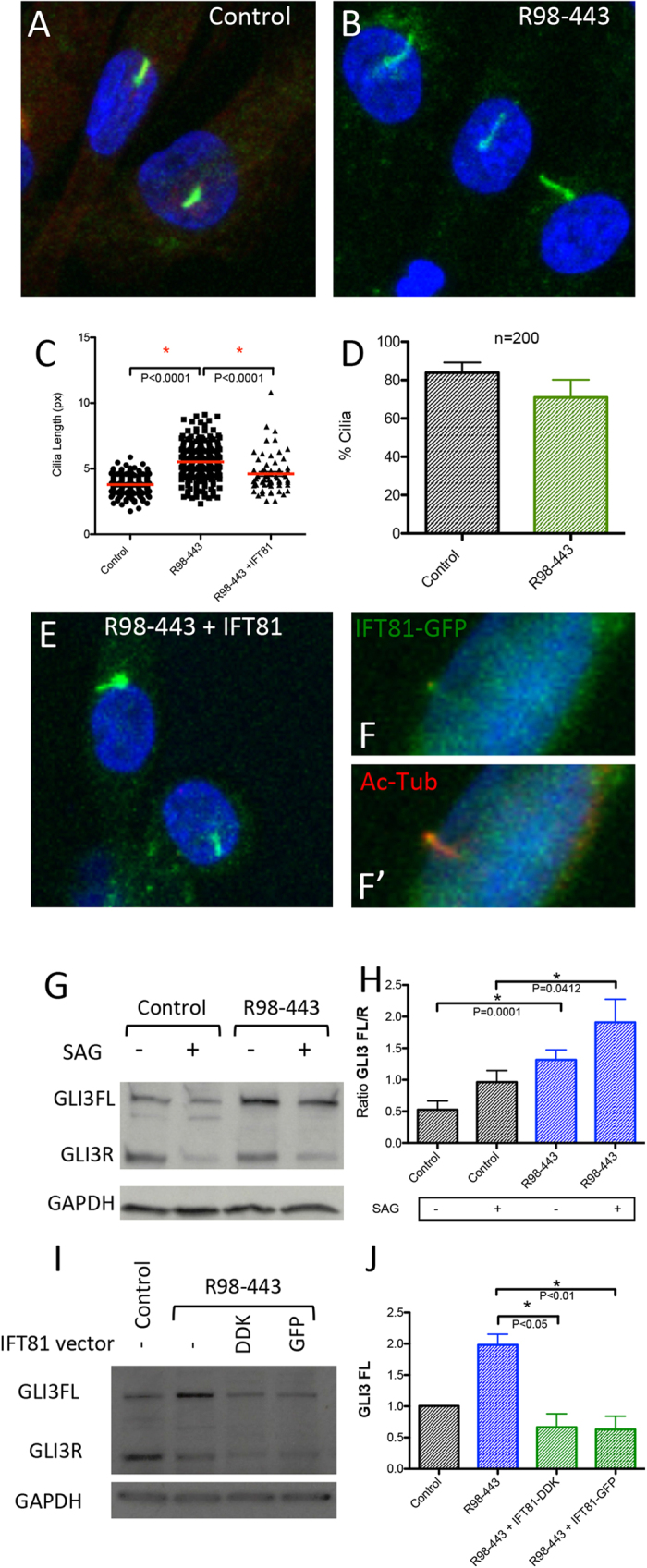
Figure 3. IFT81 mutations induce cilia defects and abnormal Hh signaling.

(A,B) ARL13B and Pericentrin staining of the centrosome and cilia in green in control and R98-443 chondrocytes. (C) Cilia length of control, R98-443 and rescued R98-443 chondrocytes with IFT81 vector showing that rescued cells partially corrected cilia length phenotype. (D) Percentage of cells with cilia in control and patient chondrocytes showing no difference in number. (E,F) Cilia staining with ARL13B and Pericentrin (both green) in rescued R98-443 chondrocytes showing average length cilia. (F,F’) demonstrate that the IFT81-GFP fusion protein co-localized with Acetyl-Tubulin in the cilia. (G,H) GLI3 levels in control and R98-443 chondrocytes with and without SAG stimulation showing altered GLI3FL/R ratios. I-J GLI3FL levels were restored to control levels with R98-443 chondrocytes rescued with vectors of IFT81 fusion protein (GFP and DDK flag).
