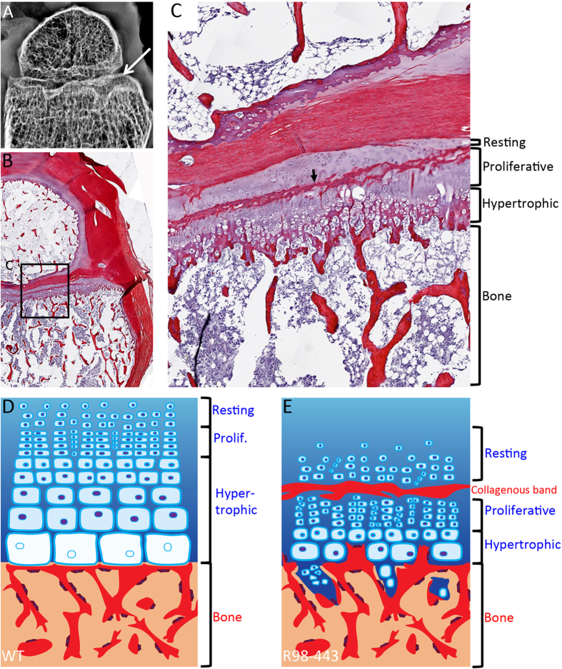
Figure 4. Growth plate defects in a patient with mutations in IFT81.
(A) X-ray of formalin fixed distal femur at 19 months of age showing irregular metaphyseal margin (arrow). (B,C) Picrosirius Red-Haematoxylin staining of same distal femur (B) magnified in (C) showing irregular hypertrophic column formation and lack of normal progressive enlargement of hypertrophic chondrocytes. Arrow points to an irregular collagenous band transecting the growth plate. (D,E) Differences between control and IFT81 defective growth plates. (D) Control. (E) R98-443. Bone is represented in red and cartilage in blue.

