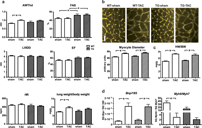
Figure 2. Response of TBC1D10C TG mice towards aortic banding (TAC)–induced hypertrophy.
(a) 8-week-old TBC1D10C TG mice underwent TAC intervention with a 27G needle or were sham operated. Echocardiography 8 weeks after surgery revealed significantly increased diastolic anterior wall thickness (AWThd) in WT TAC vs. WT sham mice, but there was no difference between WT and TG, or TG sham vs. TG TAC animals. Left ventricular inner diameter (LVIDd) was not different, and fractional area shortening (FAS) and ejection fraction (EF) were increased in TG vs. WT. (n = 9–17 mice per group; P < 0.05). Morphometry 8 weeks after the TAC intervention demonstrates increased lung weight as a sign of pulmonary congestion secondary to heart failure in TAC vs. sham operated animals. (Note: Echocardiography at 4 weeks after TAC is shown in supplemental Fig. 3). (b) WGA-stained myocardial sections were used for measurement of cardiomyocyte diameter. WT and TG mice displayed equally induced cellular hypertrophy. Scale bar: 20 μm (n = 7–9). (c) Heart weight/body weight was significantly increased after 8 weeks of TAC. (n = 7–15). (d) Real-time RT-PCR revealed a significant increase in Bnp expression and a decrease of Myh6/Myh7 after 8 weeks of TAC, but no significant differences between WT and TG (n = 7–13). (SQ: template starting quantity.)

