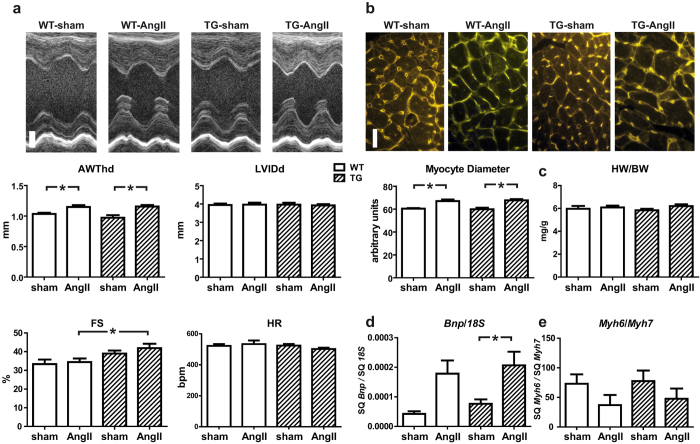
Figure 3. Response of TBC1D10C TG mice towards angiotensin II-induced hypertrophy.
(a) WT and TBC1D10C TG mice underwent chronic angiotensin II (AngII) infusion using micro-osmotic pumps. Original echocardiographic recordings after 2 weeks of AngII treatment (top; scale bar: 1 mm) and cumulative data (below). Diastolic anterior wall thickness (AWThd) was significantly increased after AngII treatment, whereas left ventricular inner diameter (LVIDd) was unchanged. Fractional shortening (FS) was increased in TG mice after AngII treatment (WT sham: n = 6; WT AngII: n = 9; TG sham: n = 6; TG AngII: n = 9). (b) Wheat germ agglutinin (WGA) stain of cardiac sections revealed a significant increase in cardiomyocyte diameter in AngII-treated mice, but no differences between WT and TG (n = 6–8 mice, 100 cells each, P < 0.05, one-way ANOVA). Scale bar: 20 μm. (c) Heart weight to body weight ratio was not increased after 2 weeks of AngII treatment (WT sham: n = 6; WT AngII: n = 9; TG sham: n = 6; TG AngII: n = 9). (d) Quantitative real-time RT-PCR of Bnp mRNA expression normalized to 18S RNA and the ratio of α- (Myh6) to β- (Myh7) myosin heavy chain gene transcript levels (WT sham: n = 6; WT AngII: n = 9; TG sham: n = 6; TG AngII: n = 9; P < 0.05; one-way ANOVA). (SQ: template starting quantity.)

