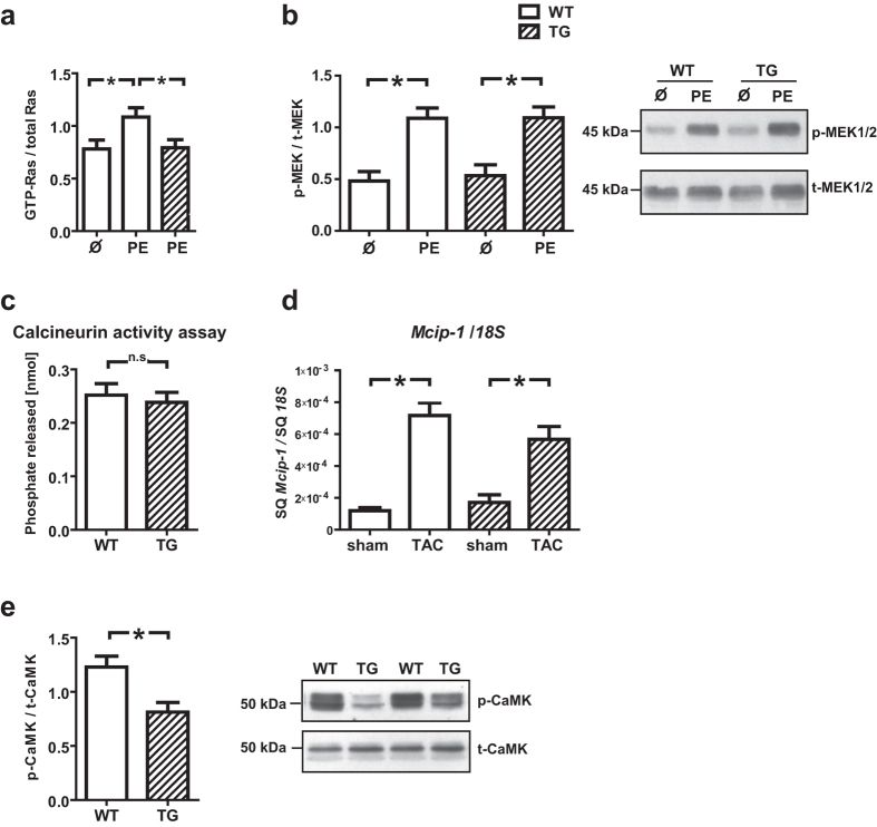
Figure 4. Examination of Ras/MAPK, CaMKII and CaN activity.
(a) Ras activity was significantly reduced in TBC1D10C TG vs. WT mice (after phenylephrine (PE) stimulation) (WT, uninjected: n = 4; WT + PE: n = 14; TG + PE: n = 15; P < 0.05; Student’s t-test), whereas (b) p-MEK1/2/t-MEK1/2 levels were not significantly diminished in PE stimulated TG vs. PE stimulated WT mice (WT, uninjected: n = 3; WT + PE: n = 15; TG, uninjected: n = 3; TG + PE: n = 15,n.s.; Student’s t-test). (c) Calcineurin (CaN) phosphatase activity was not different between WT and TG mice (n = 6 per group), suggesting CaN-independent signal pathways may be more important for the effects of TBC1D10C in vivo. (d) Mcip-1 (Modulatory calcineurin-interacting protein 1) mRNA expression, a marker for CaN activity, was not significantly changed in TG mice whereas (e) CaMKII (T286) phosphorylation was significantly diminished in TG mice (TG: n = 8; WT: n = 10; P < 0.01; Student’s t-test). Full length blots of the cropped membranes in Fig. 4b and e are displayed in Supplementary Figure 6.

