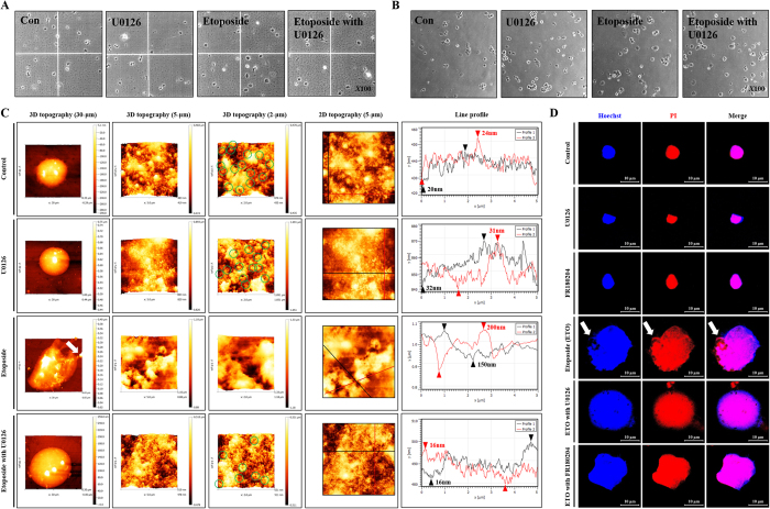
Figure 5. Etoposide induced nuclear envelope ruptures through ERK activation.
HK-2 cells were pre-treated with U0126 (20 μM) or FR180204 (10 μM) for 1 hour followed by treatment with etoposide (50 μM) for 48 hours. Nuclei were extracted from the cells using NE-PER®nuclear and cytoplasmic extraction reagents. (A) Nuclear morphological changes were measured using a hematology analyzer and detected by microscopy (original magnification X100). (B) Nuclei were extracted, seeded and attached on culture dish for 15 minutes, where morphological changes such as nucleus swelling were measured using microscopy (original magnification X100). (C) Topography of NE was measured using an AFM probe system. Images shown are representative of 3-D topography at 30, 10, and 2 μm scales. (D) Nuclear extracts were seeded on coverslips for 15 min and stained with nucleus targeting dyes such as Hoechst 33258 (blue color) and propidium iodide (red color) that were detected using confocal microscopy. Arrows indicate the DNA leakage.

