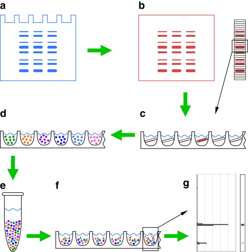
Figure 1. Bead-based western blot (DigiWest) workflow.
(a) Protein separation by gel electrophoresis, usually SDS-PAGE. (b) Blotting of proteins to membrane and biotinylation of immobilized proteins directly on the membrane. (c) The cutting of sample lanes into 96 stripes to generate 96 molecular weight fractions immobilized on the membrane; elution of the proteins in 96-well plates. (d) Loading of biotinylated proteins onto 96 distinct Neutravidin-coated magnetic Luminex bead-sets. (e) Pooling into bead pools and reconstitution of the initial sample lane. (f) Immunoassay: aliquots of the generated bead pool (<0.5%) are incubated with western blot antibodies overnight before PE-labelled secondary antibodies are added for signal generation. (g) Readout using a Luminex instrument, reconstitution of the initial lane and data analysis.

