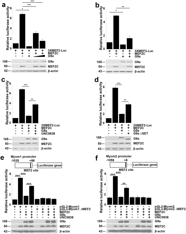
Figure 4. G9a represses MEF2C transcriptional activity.
(a,b) Luciferase assays were performed using 100ng of 3XMEF2-Luc reporter in (a) 10T1/2 fibroblasts or (b) C2C12 myoblasts co-transfected with 100 ng Flag-MEF2C in the presence or absence of 50 and 100 ng Flag-G9a as indicated. Luciferase activity was measured 24 hr after transfection. Lysates were analyzed by western blot for exogenous G9a and MEF2C expression. (c) 100 ng 3XMEF2-Luc reporter was transfected in 10T1/2 fibroblasts with 100 ng Flag-MEF2C and 100 ng Flag-G9a. Cells were recovered for 24 hr after transfection and then treated with UNC0638 for 48 hr, and luciferase activity was measured after treatment. (d) 100 ng 3XMEF2-Luc reporter was transfected with 100 ng Flag-MEF2C and 100 ng Flag-G9a or 200 ng Flag-G9a∆SET. Luciferase activity was measured 24 hr after transfection. Western blot was done to analyze expression of G9a and MEF2C in the lysates. Error bars indicate mean ± standard error of n = 3. (e,f) Schematic diagram depicting Myom1 (pGL3-Myom1) and Myom2 (pGL3-Myom2) promoter luciferase constructs. The MEF2 binding site within the promoters is indicated and was mutated (pGL3-Myom1∆MEF2 and pGL3-Myom2∆MEF2 respectively). Luciferase assays were done using pGL3-Myom1 and pGL3-Myom1∆MEF2 (e), and pGL3-Myom2 and pGL3-Myom2∆MEF2 (f) in 10T1/2 fibroblasts co-transfected with MEF2C in the presence or absence of G9a, treated with UNC0638 or DMSO for 48 hr. Western blot shows expression of exogenous G9a and MEF2C. Error bars indicate mean ± standard deviation. The data are representative of three independent experiments.

